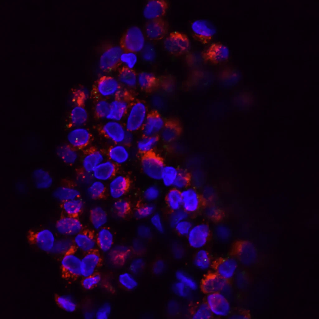
Breast cancer is a type of cancer that develops in the cells of the breast. It forms when there is an uncontrolled growth of abnormal cells in the breast tissue. These abnormal cells can form a lump or mass, and can also spread to other parts of the body through the blood or lymph system. Breast cancer is the most common cancer in women in the United States. It is also known as carcinoma breast cancer because it begins in the cells that make up the breast tissue called ductal or lobular epithelium.
A research conducted by “Cancer Research UK” shows that regular mammograms can reduce breast cancer deaths by 25 percent. Breast cancer screening is strongly recommended for women over 50 years’ old who have a life expectancy of 10 years or more and are at increased risk due to genetic factors, family history of breast cancer or other factors such as high body weight.
SYMPTOMS OF BREAST CANCER
Breast cancer can cause a variety of symptoms, but the most common symptoms are:
Lumps or thickening in the breast or underarm area
A lump or thickening in the breast or underarm area is the most common symptom of breast cancer. It may feel hard, irregular in shape, and may be painless. It may feel hard, irregular in shape, and may be painless. It’s important to get checked out by a doctor if you notice any of these symptoms.
Most cases are not cancer, but rather cysts or breast infections – both of which can be easily treated with medication and do not require treatment from a surgeon. In some rare cases, the lump is actually cancerous and will require surgery to remove the tumor.
Changes in the size or shape of the breast
Breast cancer can cause the breast to change shape or size. The breast may become larger, smaller, or may have an unusual shape.
Dimpling or puckering of the skin
Breast cancer can cause the skin of the breast to become dimpled or puckered, like the skin of an orange. It is characterized by a noticeable change in the appearance of the skin on the breast, causing it to appear like it’s been pinched or dimpled. This type of skin change can be caused by a tumor that is growing beneath the skin and pulling it inward.
This can cause the skin to appear dimpled, puckered, or like an orange peel. The dimpling or puckering can also be accompanied by changes in the size or shape of the breast, redness, or thickening of the skin. It’s important to note that not all cases of dimpling or puckering of the skin are caused by breast cancer. Other conditions such as fibrocystic breast changes, infection, or injury can also cause this type of skin change.
Nipple Changes
Breast cancer can cause changes in the nipple such as redness, scaliness, or discharge from the nipple. The nipple may also become inverted or change position. It is important to note that not all nipple changes are caused by breast cancer. Hormonal changes, infection, or injury can also cause changes in the nipple. However, if you notice any changes in your nipple or breast area, it is important to consult with a healthcare professional for proper diagnosis and treatment.
Swelling or discomfort in the Breast or Armpit
Breast cancer can cause swelling or discomfort in the breast or armpit. Another possible cause of breast swelling is infection. Mastitis, an infection of the breast tissue, can cause breast pain, swelling, and redness. It is most common in women who are breastfeeding.
Hormonal changes in the body can also cause breast swelling and discomfort. This can happen during the menstrual cycle, pregnancy, or menopause.
Skin irritation or Rash
Breast cancer can cause the skin of the breast to become red, irritated, or itchy. This is called carcinoma ex pleomorphic leuakoplakia (CEL). It does not always mean that there are cancer cells present, but it’s best to be checked out by a doctor just in case.
It’s important to note that breast cancer may not cause any symptoms in its early stages. It’s important for women to be aware of their own breast tissue and to report any changes to their healthcare provider.
It is also important to note that, these symptoms can also be caused by non-cancerous conditions such as cysts or infection. So, If you notice any changes in your breast, it is important to see your healthcare provider to determine the cause.

SELF EXAMINATION OF BREAST CANCER
Self-examination of the breasts, also known as breast self-examination (BSE), is a method of detecting breast cancer early by regularly checking the breasts for any changes or abnormalities. It can be done at home, and it’s an important part of breast health for women of all ages.
Here are the steps for performing a breast self-examination:
1. Stand in front of a mirror with your arms at your sides and look for any changes in the shape, size or symmetry of your breasts.
2. Raise your arms above your head and look for any changes in shape or size.
3. While standing, place your hands on your hips and press down firmly to flex your chest muscles. Look for any dimpling or puckering of the skin.
4. While lying down, use the pads of your fingers to feel for lumps or abnormalities in your breasts. Begin by feeling the breast tissue closest to your underarm and work in small circles, moving from the outside to the center of the breast. Be sure to feel all the tissue from your collarbone to your breastbone and from your armpit to your cleavage.
5. Repeat the process on your other breast.
It is important to keep in mind that breast self-examination is not a substitute for regular mammograms and clinical breast exams. Women should also consult with their healthcare professional about the appropriate schedule for mammograms and clinical breast exams.
PHYSICAL EXAMINATION OF BREAST CANCER
Breast cancer can be diagnosed through physical examination methods, including:
Physical examination: A doctor will examine the breast for any lumps or changes in shape or size. A physical examination of the breast is one of the methods used to screen for and diagnose breast cancer. The examination is typically done by a doctor or a nurse practitioner, and includes the following steps:
Inspection: The doctor will visually inspect the breast, looking for any changes in shape, size, or symmetry. They will also check for any dimpling or puckering of the skin, which can be a sign of breast cancer.
Palpation: The doctor will use their hands to feel for any lumps or masses in the breast tissue. They will also check the lymph nodes under the arm and above the collarbone to see if there is any swelling, which can be a sign of cancer that has spread.
Nipple examination: The doctor will examine the nipples for any discharge, retraction (turning inward), or changes in texture.
Additional tests: If a lump or abnormality is found during the physical examination, the doctor may recommend additional tests such as a mammogram or ultrasound to further evaluate the area.
It’s important to note that a physical examination of the breast is not a perfect method for detecting breast cancer, as some lumps or abnormalities may not be felt during the examination. Therefore, it’s important for women to also undergo regular mammography screening as per their doctor’s recommendation and to be familiar with their own breasts so that they can recognize any changes and report them to their doctor.

DIAGNOSIS OF BREAST CANCER
Mammography
This is a specialized X-ray that can detect small tumors or abnormalities in the breast tissue. A mammogram is often used as the first line of screening for breast cancer, especially in women over 50. Breast cancer can also be classified according to its stage, grade and hormone receptor status. The American Cancer Society recommends that women get regular mammograms starting at age 40.
A mammogram is a special X-ray of the breast used for screening and diagnosis of abnormalities in the breast tissue. It is important for women to have routine mammograms because they are able to detect breast cancer early on when it is most treatable. There are different types of mammograms:
DIGITAL MAMMOGRAPHY
In this type of mammography, the X-ray images are captured digitally, rather than on film. This allows for more detailed images and makes it easier to spot small tumors or abnormalities. It also allows for the images to be magnified and enhanced, making it easier for radiologists to identify potential abnormalities.
3D MAMMORAPHY (TOMOSYNTHESIS)
This type of mammography uses a low-dose X-ray and a computer to create detailed, three-dimensional images of the breast tissue. The images are taken in “slices” that can be viewed individually or in a series, which makes it easier for radiologists to identify abnormalities, especially in women with dense breast tissue.
MAGNETIC RESONANCE IMAGING (MRI)
This type of mammography uses a combination of a magnetic field and radio waves to create detailed images of the breast tissue. It is often used in women with a higher risk of breast cancer or for women who have already been diagnosed with the disease.
CONTRAST-ENHANCED MAMMOGRAHY
This type of mammography uses a contrast agent (such as a dye) to make abnormal areas of the breast tissue more visible on the images. It is usually used in conjunction with traditional mammography or MRI.
It’s important to note that mammography is not a perfect screening tool and it can miss some cancers and give false-positive results. It’s recommended that women should discuss with their doctor about the best screening schedule and type of mammography for their individual needs.
Ultrasound
This test uses high-frequency sound waves to create images of the breast tissue. It can be used to determine if a lump is a solid mass or a fluid-filled cyst. Ultrasound is a non-invasive imaging test that uses high-frequency sound waves to create images of the breast tissue. It is often used in combination with mammography to evaluate breast lumps or abnormalities that have been identified.
The process for an ultrasound of the breast typically involves the following steps:
Preparation: You will be asked to remove any clothing or jewelry that may interfere with the imaging process. You will then be given a gown to wear for the test.
Positioning: You will lie on your back on the examination table with your arm behind your head. A small amount of gel will be applied to the breast to help with the transmission of the sound waves.
Imaging: The ultrasound technician will use a hand-held device called a transducer to take images of the breast tissue. The transducer sends out sound waves and then listens for the echoes as they bounce back. These echoes are then used to create images of the breast tissue. The technician will move the transducer over the breast to get images from different angles.
Duration: The entire process usually takes around 20-30 minutes.
Results: The images will be analyzed by a radiologist and the results will be sent to your doctor. If an abnormal area is found, your doctor may recommend a biopsy to confirm the diagnosis.
It’s important to note that, ultrasound is not as sensitive as mammography in detecting breast cancer, particularly in women with dense breast tissue. Therefore, it is typically used in addition to mammography, rather than as a standalone test.
Additionally, ultrasound is not recommended as a screening tool for breast cancer, it’s mostly used as a diagnostic tool after a lump or other abnormality is detected through self-examination, clinical examination or mammography.

Biopsy
Biopsy is the removal of a small piece of tissue from the breast for examination under a microscope. A biopsy is the only way to confirm a diagnosis of breast cancer. A biopsy is a procedure in which a small sample of breast tissue is removed and examined under a microscope to determine if cancer is present. There are several different types of biopsy procedures that may be used to diagnose breast cancer, including:
Fine-Needle Aspiration (FNA) Biopsy
During this procedure, a thin needle is inserted into the breast to remove a small sample of tissue. The tissue is then examined under a microscope to see if cancer cells are present. This procedure is typically used for small or deep-seated lumps that are not visible on a mammogram.
Core Needle Biopsy
During this procedure, a larger needle is inserted into the breast to remove a small cylinder of tissue. The tissue is then examined under a microscope to see if cancer cells are present. This procedure is typically used for larger lumps that can be felt during a physical examination or seen on a mammogram.
Surgical Biopsy
During this procedure, a small incision is made in the breast and a small piece of tissue is removed. The tissue is then examined under a microscope to see if cancer cells are present. This procedure is typically used for larger lumps that can be felt during a physical examination or seen on a mammogram, or for lumps that cannot be removed by fine-needle or core needle biopsy.
Image-Guided Biopsy
This type of biopsy uses imaging techniques like ultrasound, MRI or CT scan to guide the needle to the abnormal area. This procedure can be used in case of non-palpable lumps or abnormalities not seen on mammograms.
The specific type of biopsy will depend on the size, location, and characteristics of the lump or abnormality. The biopsy procedure usually takes around 30 minutes to an hour and is usually done on an outpatient basis. It’s important to note that biopsy is the only definitive way to confirm a diagnosis of breast cancer and that biopsy results can take several days to be available.
Blood Tests
Certain biomarkers such as CA15-3, CA27.29, and CEA are elevated in certain types of breast cancer and can be useful in monitoring the disease. There are several blood tests that may be used to help diagnose breast cancer, but the most common is called a tumor marker test. This test measures the levels of certain substances, such as hormones or proteins, in the blood that are often elevated in people with breast cancer. Some examples of tumor markers for breast cancer include CA 15-3, CEA, and HER2/neu.
The process for a blood test for breast cancer typically involves a healthcare professional drawing a sample of blood from the patient, usually from a vein in the arm. The blood sample is then sent to a lab for analysis, where it is tested for the presence of tumor markers.
It’s important to note that a positive result on a blood test for breast cancer does not necessarily mean that the person has cancer. Elevated levels of tumor markers can also be caused by other conditions, such as benign breast disease or non-cancerous tumors. Therefore, a positive result on a blood test is typically followed by further testing, such as a biopsy, to confirm the diagnosis.
Additionally, some blood tests for breast cancer are used for monitoring of breast cancer patients who are already diagnosed, to evaluate the progression of the disease and the effectiveness of treatment. It’s also important to note that, blood tests are not a primary diagnostic tool for breast cancer, and are typically used in conjunction with other diagnostic tests such as mammography, ultrasound, and biopsy.
SOME NOTES TO REMEMBER
In conclusion, breast cancer is a serious condition that can cause various symptoms, including swelling or discomfort in the breast or armpit, nipple changes, dimpling or puckering of the skin, and changes in the size or shape of the breast. It’s important to be aware of these symptoms and to consult with a healthcare professional if any of them are noticed. Early detection is key to effectively treating breast cancer, and a combination of methods such as physical examination, mammography, and ultrasound may be used to diagnose the condition. If breast cancer is diagnosed, treatment options such as surgery, radiation therapy, and chemotherapy are more likely to be effective if the cancer is caught early.